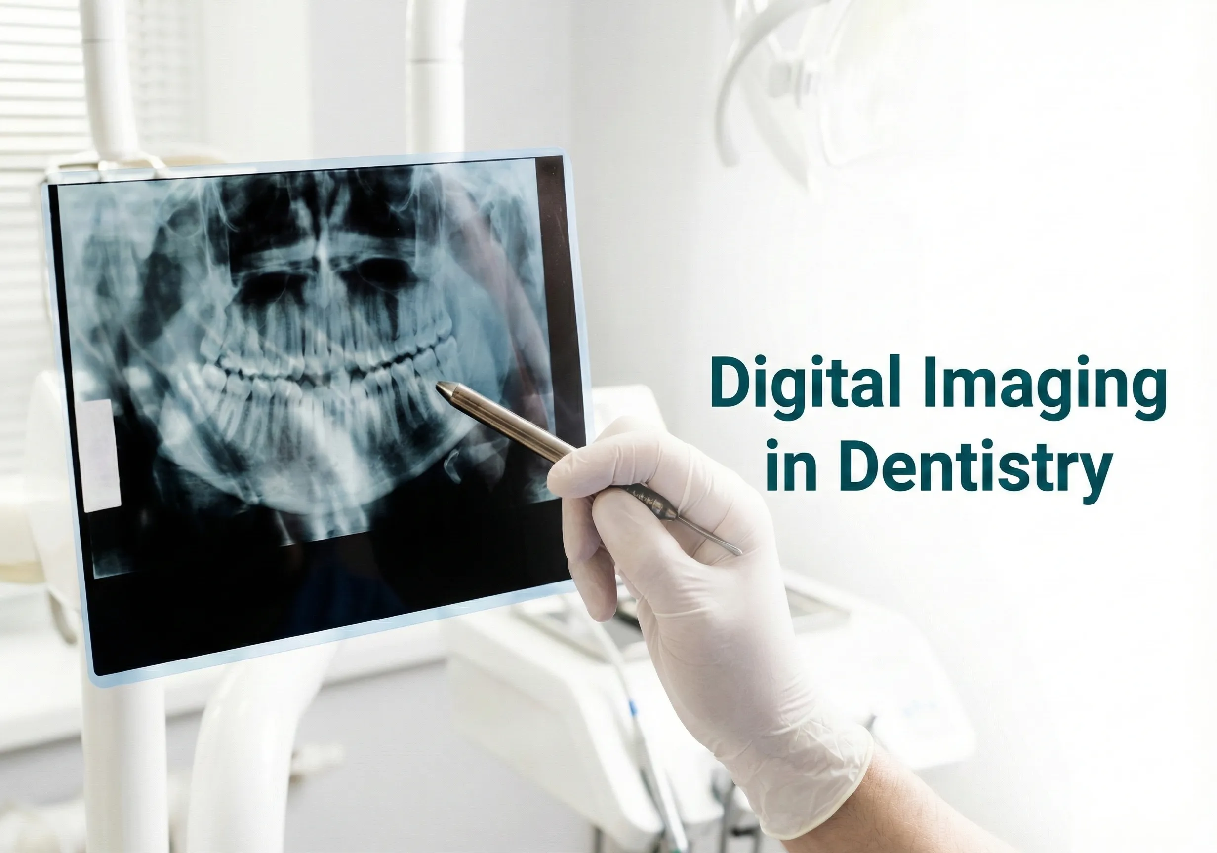
At Dentist in Winston Salem, accurate diagnosis is the foundation of effective dental care. Digital imaging is a modern method of dental radiography that has transformed how dentists diagnose oral conditions.
By replacing traditional film-based X-rays, digital radiography allows faster, clearer, and more efficient imaging—benefiting both patients and clinicians.
Dental imaging uses X-rays to capture detailed images of teeth, bones, and surrounding structures.
Many dental conditions—such as cavities, periodontal disease, or bone loss—are difficult to detect through clinical examination alone.
Digital X-ray images allow dentists to identify these conditions early, saving time and improving treatment planning.
At Dentist in Winston Salem, digital imaging helps us diagnose conditions accurately and explain treatment needs clearly to our patients.
X-rays were first discovered in 1895, and conventional film radiography was widely used for decades.
Since the mid-1980s, digital radiography has largely replaced traditional screen-film methods.
Digital imaging uses solid-state sensors to capture images, which are instantly displayed and stored on a computer.
This advancement has significantly improved diagnostic precision and workflow efficiency.
Clinical advantages include:
Digital imaging is also widely used in forensic dentistry for age estimation, dental identification, postmortem examinations, and legal documentation.
Intraoral radiographs are taken from inside the mouth and provide detailed views of teeth and supporting structures.
Common intraoral techniques include:
Extraoral radiographs are taken from outside the mouth and are especially useful when mouth opening is restricted due to trauma or pathology.
Common extraoral techniques include:
Digital radiography requires significantly less radiation exposure compared to traditional E-speed film.
One digital image can be adjusted for multiple diagnostic purposes—lighter images for bone evaluation and darker images for caries detection—without retaking X-rays.
This makes digital imaging both safe and efficient for patients.
At Dentist in Winston Salem, we use digital imaging to ensure precise diagnosis, safer imaging practices, and transparent communication with our patients.
Advanced imaging allows us to detect problems early and tailor treatment plans effectively.
If you need an accurate dental diagnosis or want to understand your oral health better, schedule your appointment with Dentist in Winston Salem today.
We’re here to help you make informed decisions about your dental health.
Digital radiography requires significantly less radiation exposure than traditional E-speed film, which makes it a safer option for dental imaging. A single digital image can also be adjusted afterwards for different diagnostic purposes, lighter for evaluating bone and darker for detecting caries, so fewer repeat exposures are needed. Images are taken only when clinically indicated.
Digital imaging is a modern form of dental radiography that uses solid-state sensors instead of film to capture X-ray images, which then appear instantly on a computer and are stored electronically. It has largely replaced screen-film methods since the mid-1980s. The result is faster, clearer imaging and better diagnostic precision for problems an examination alone cannot reveal.
Periapical X-rays show an entire tooth from crown to root along with the surrounding bone, which helps detect apical inflammation, fractures, cysts, unerupted teeth, and periodontal problems. Bitewing X-rays capture upper and lower teeth together in one view and are most effective for finding decay between teeth and assessing bone levels. Both are intraoral techniques.
Many dental conditions cannot be seen during a visual examination. Decay between teeth, bone loss, infection at a root tip, impacted teeth, and cysts all sit below the surface. X-ray images let a dentist find these problems early, when treatment is simpler, and they also make it easier to explain why a particular treatment is being recommended.
A panoramic image, also called an OPG, is an extraoral X-ray that provides a full-mouth overview in a single picture. It is helpful for evaluating jaw fractures, TMJ conditions, impacted teeth, and the overall structures of the jaws. Extraoral imaging is also useful when mouth opening is restricted because of trauma or pathology.
Tell us a little about what you need — we'll be in touch shortly.
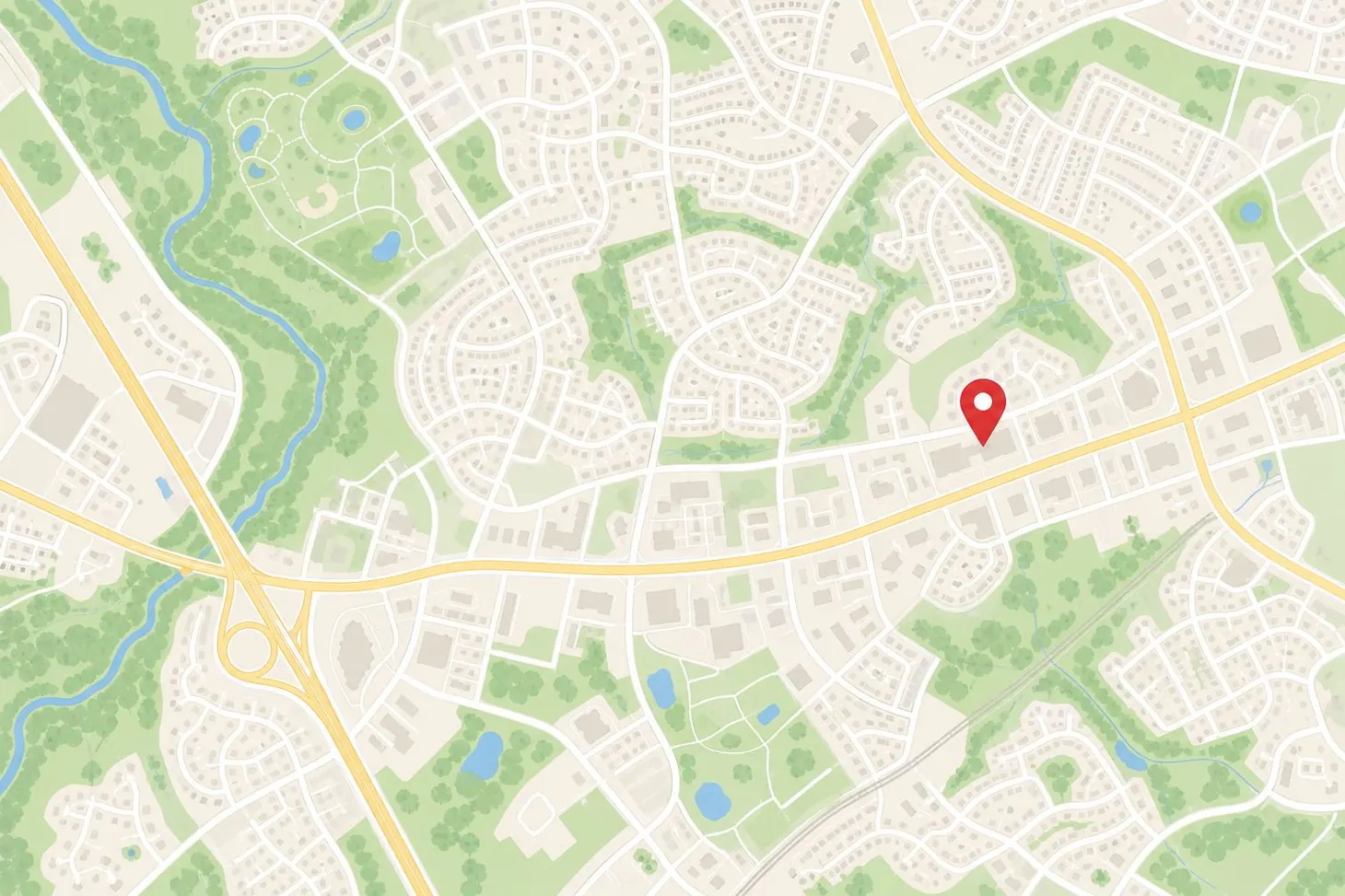
3210 Silas Creek Pkwy, Suite-4, Winston-Salem, NC, 27103, United States

Hi! I'm Tooth fairy! Your AI Assistant Ask me anything